Get to know your horse's hoof, and what a healthy one should look like! Healthy horses, Horse

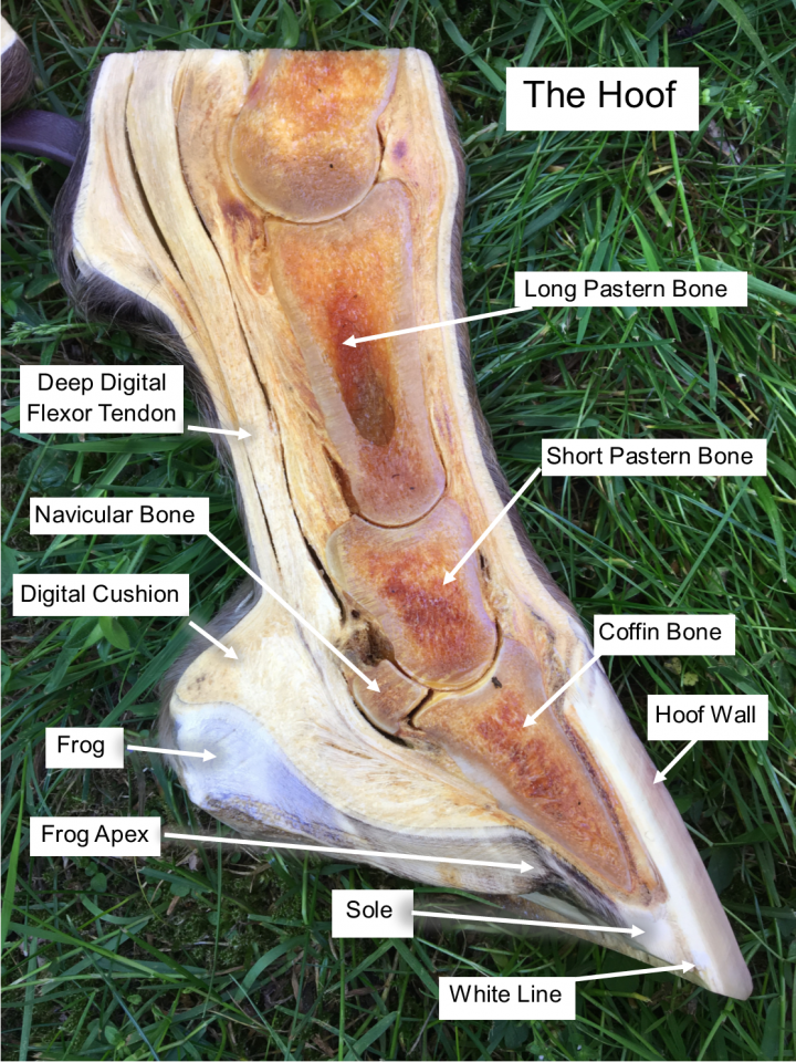
There are two and a bit bones inside the hoof. The Pedal bone, the Navicular bone and the bottom part of the Short Pastern bone. Pedal Bone. The large bone inside the hoof capsule is known as the Pedal bone or Coffin bone. Its shape provides a framework for the shape of the hoof capsule itself.
Horse Foot Anatomy Diagram Quizlet

Horse Hoof Anatomy, Part 1. Learn how the bones and soft tissues in a horse's hoof work together and impact soundness. When blood flow within the hoof to the coffin bone is compromised for long.
Horse care, Equine veterinary, Horse anatomy

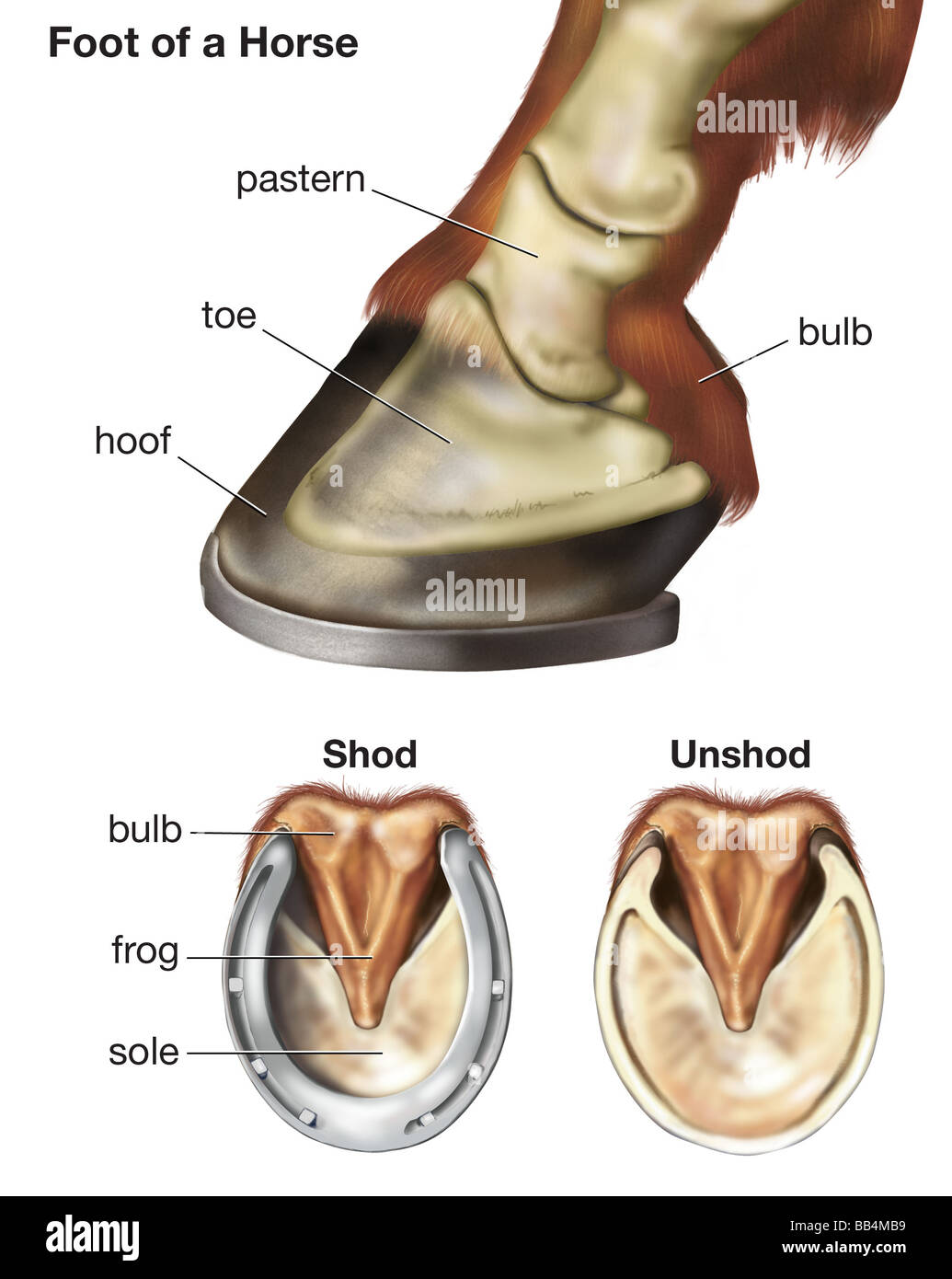
Department of Veterinary Anatomy. College of Veterinary Medicine. A horse's hoof is composed of the wall, sole and frog. The wall is simply that part of the hoof that is visible when the horse is standing. It covers the front and sides of the third phalanx, or coffin bone. The wall is made up of the toe (front), quarters (sides) and heel.
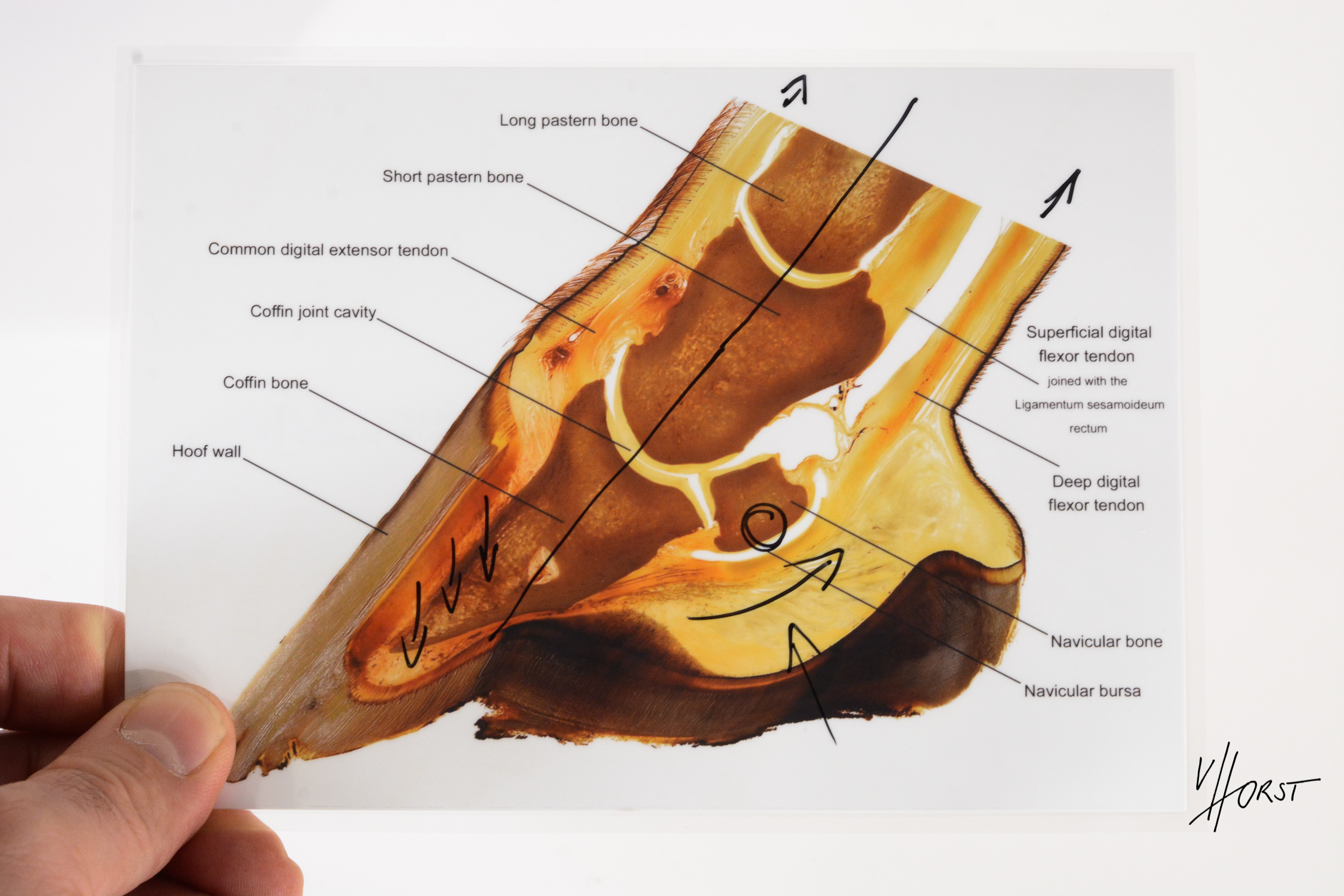
Ligaments and tendons of the equine hoof. Horse anatomy, Horse health, Equine veterinary

The equine foot is a mechanical marvel of joints, cartilage, ligaments, tendons, blood vessels and bone encased within a horny capsule that is vital to the horse. Dr. Stephen E. O'Grady of Keswick, Virginia, has devoted his professional life to its study, both as a veterinarian and farrier. He's an acknowledged worldwide expert on equine.
Horse anatomy by Herman Dittrich lower leg and hoof Shoestring Stable Horse drawings

The Anatomy Of A Horse's Foot. When we talk about hooves, we picture the entire anatomy of a horse's foot. However, the hoof is only one part of the larger anatomy. There are many parts of a horse's foot anatomy, including: Periople: The periople covers the coronary band at the top of the hoof structure where the hoof meets the coat. The.
Labeled Hoof Diagram Cavallo Hoof Boots Horse Boots, Hoof Boots, Saddle Pads
1. Frog - This is a rubbery wedge shaped structure positioned between the bars. 2. Bars - There are two bars on each hoof. They are on either side of the frog. 3. Sole - The sole covers the bottom of foot. 4. White line - This is actually inter-connected lamina that you can see.
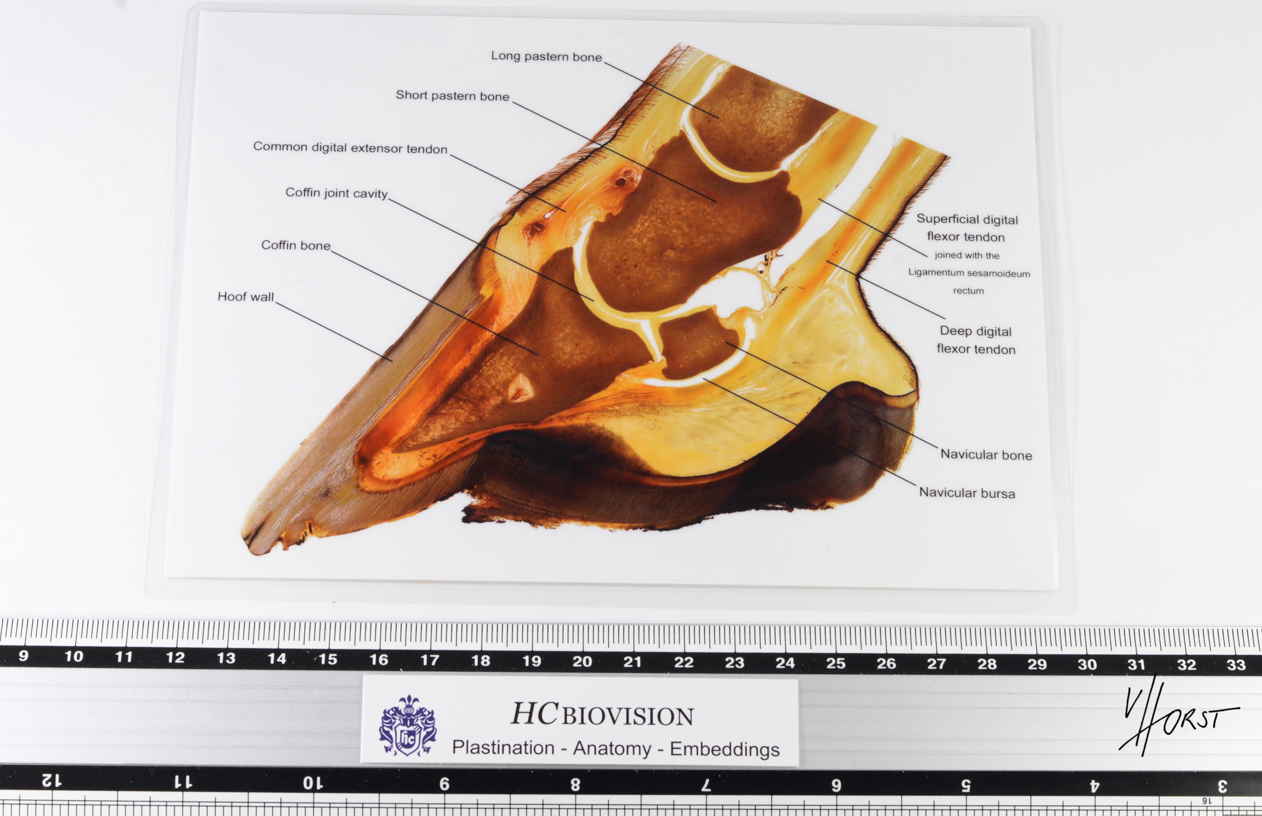
Laminated hoof anatomy chart print Plastination Anatomy Embedding
This article provides an overview of foot anatomy and physiology, with a focus on fundamental knowledge. The foot is defined as the epidermal hoof capsule and all structures enveloped by the capsule. The anatomy is described using terminology published in Nomina Anatomica Veterinaria.
Horse anatomy, Hoof care, Horses

General Anatomy Of The Hoof. Let's start by looking at the following diagram, which shows basic outer hoof anatomy. Knowing these words and the areas they refer to on a horse's hooves will allow you to better understand your resident's mobility, provide better care, and communicate more effectively with an equine veterinarian and farrier.
Vitals & Anatomy Horse Side Vet Guide Horse anatomy, Horses, Anatomy

Inflammation of the sensitive laminae which attach the hoof capsule to the fleshy portion of the foot. In laminitis, the blood flow to the laminae is affected, resulting in inflammation and swelling in the tissues within the hoof, and severe pain. As the laminae are starved of oxygen and nutrient rich blood, the cells become damaged.
Leg anatomy Horse health, Horse care, Horse anatomy

Enter search terms to find related medical topics, multimedia and more. Advanced Search: Use " " for exact phrases. For example: "canine influenza"
Anatomy of lower horse leg/foot. Horses, Horse anatomy, Horse health

Hoof Anatomy: What Horse Hooves are Made of. The equine hoof is a great example of Mother Nature's engineering capabilities. Consider the size and weight of a horse relative to the size of a hoof, and how fast horses can run or how high they can jump; it's amazing how so much is supported by so little. A horse's hooves play a key role in.
Guide to a Horse's Foot r/coolguides
Discover the anatomy of a horse's foot, including its external and internal structures, functions, common problems, and essential care techniques for maintaining healthy hooves. External Structures of a Horse's Foot Hoof Wall. The hoof wall is the tough, outer layer that surrounds and protects the internal structures of a horse's foot.
Laminated hoof anatomy chart print Plastination Anatomy Embedding
Hoof anatomy. The equine hoof is a unique structure which bears a lot of weight over a small surface area. The term 'no foot, no horse' is extremely important as issues with the hoof can cause major health and movement issues. The hoof is a complex makeup of structures built to withstand tremendous forces, adapt to varying terrains and.
tobreedornottobreed From Dr. Deb Principles of Equine Orthopedics Part 1 Vet

Horse hoof. Barefoot hoof, lateral view. (1) Coronet band, (2) walls, (3) toe, (4) quarter, (5) heel, (6) bulb, (7) P2 (small pastern) A horse hoof is the lower extremity of each leg of a horse, the part that makes contact with the ground and carries the weight of the animal. It is both hard and flexible. It is a complex structure surrounding.
Foot of a horse showing the position of skeletal bones, as well as the difference between a shod
The "foot" of ungulates is generally defined as the epidermal hoof capsule and all the tissues and structures enveloped by the capsule, including dermis, subcutaneous tissue, neurovascular tissues, bone, synovial spaces, tendon, ligament, and cartilage. The tremendous weight-bearing forces transmitted through the 4 digits of the horse are.
Hoof Care Tips and Anatomy

Horse hoof anatomy. First, I would like to introduce the different parts of a horse hoof anatomy. You will mainly find the three parts - wall, sole, and frog in a horse hoof.
.